What images can’t tell you, and what your body already knows.
For athletes, runners, and active adults. 5min read
You pulled something during your last long run. Your shoulder has been nagging you for weeks. You’re dealing with low back pain that showed up out of nowhere. The instinct is understandable: I need a scan. I need to see what’s wrong.
It makes sense. We live in a culture that trusts pictures. If we can see it, we can fix it. But when it comes to the kind of musculoskeletal pain that sidelines athletes, runners, and active adults, imaging tells a surprisingly incomplete story. And sometimes, it sends people down a road they didn’t need to travel.
The MRI Trap
Here’s a fact that might surprise you: getting an MRI for low back pain is one of the leading predictors of eventually having back surgery. Not because the surgery was necessarily needed, but because of what happens after the image is taken.
A 2008 study found that nearly 1 in 3 back pain patients should not receive an MRI because it may lead to unnecessary spinal surgery. Clinical guidelines in the Journal of Athletic Training now explicitly state that clinicians should refrain from routine, immediate lumbar imaging for patients with nonspecific, acute, or subacute low back pain when there are no signs of a serious underlying condition.
The problem isn’t the technology itself. MRIs are powerful tools when used at the right time, for the right reasons. The problem is what happens when a scan becomes the first stop instead of a last resort. An MRI report can describe findings like “disc bulge,” “degenerative changes,” or “mild stenosis” in perfectly healthy, pain-free people. If those same findings show up in someone with pain, now they are associated with the pain and seen as a problem. However, these anatomical changes (which are as normal as a gray hair) could have been present before the onset of the pain. In other words, MRIs don’t tell us where the pain is coming from.
In addition, when a patient reads those terms attached to their own spine, anxiety rises, catastrophizing sets in, and suddenly, the back feels worse than it did before the appointment.
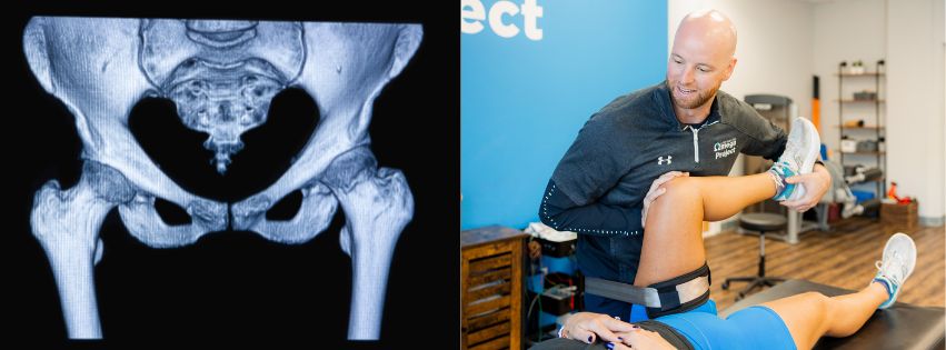
What Your Physical Therapist Sees That an MRI Cannot
An image shows structure. It cannot show movement. It cannot show how you compensate when you’re fatigued, how your hip drops when you land on your right foot, or how tension in your thoracic spine quietly loads your lower back with every stride.
This is where a skilled physical therapist, especially one trained in orthopedics and sports medicine, has an enormous advantage. Through a thorough movement assessment, manual examination, and clinical reasoning, we can identify patterns of dysfunction that no scan will ever reveal.
And here’s the part that surprises most athletes: the source of your pain is often not where the pain lives.
A runner with chronic knee pain may have a hip that isn’t loading properly. A swimmer with shoulder impingement may have a restricted thoracic spine that’s forcing the shoulder to work overtime. A desk worker with low back pain may have dormant glutes, causing the lumbar spine to compensate with every step. Imaging the painful area tells you where the fire is, not where the spark started.
For Most Injuries, PT Outperforms Surgery
Here’s something worth knowing before you schedule that imaging appointment: for the most common injuries we see (back pain, shoulder problems, and knee issues), physical therapy consistently produces outcomes that are equal to, or better than, surgery. The research on this is clear and has been for years. And yet, the pipeline from “get an MRI” to “you should consider surgery” remains well-worn, because an image almost always finds something that looks like it needs fixing.
The truth is, many of those findings were there long before your pain started — and will be there long after you recover. What gets you better isn’t addressing what showed up on the scan. It’s restoring how your body moves, loads, and absorbs force. That’s what physical therapy does. Surgery operates on a structure; PT restores a system. For the vast majority of active adults dealing with musculoskeletal pain, that’s the difference that matters.
How We Approach Your Care at Omega Project PT
Our therapists bring backgrounds in orthopedics, sports medicine, exercise science, and rehabilitation. That foundation shapes how we think about injury from the very first visit. We’re not looking for a diagnosis on a film; we’re looking for the root cause of your movement problem.
- Root cause analysis: We identify the origin of dysfunction, which is often not at the site of pain.
- Movement-based diagnosis: We assess your mobility to determine whether you have flexibility deficits or strength/stability deficits in the area of the injury and in surrounding joints. We relate these deficits to the biomechanics and cause of the dysfunction.
- Injury prevention: We don’t just resolve your current pain; we make sure you come back stronger by addressing the deficits that caused the injury.
When is Imaging the Right Call
None of this means imaging is never appropriate. If your physical therapist suspects a fracture, a serious structural injury, or a condition that warrants medical management, you’ll be referred immediately. Red flags are taken seriously. Our goal isn’t to avoid imaging at all costs — it’s to make sure imaging is used purposefully, when it will actually change the course of your care.
In most cases, for athletes and active adults dealing with pain from overuse, poor mechanics, or acute soft tissue injury, the most powerful diagnostic tool isn’t an MRI machine. It’s an expert set of hands, trained eyes, and a movement screen that tells us what your body is actually doing.
If you’ve been waiting on imaging before seeking help, you don’t have to. The sooner we understand how you move, the sooner we can get you back to doing what you love (without the waiting room, the contrast dye, and the alarm that comes from reading a report written for radiologists, not athletes).
Schedule Today!